Tumors are living ecosystems.
The Davies Cancer Lab develops living tissue models, time-lapse imaging, and computational approaches to understand how cancer cells respond to their surroundings across space and time— to reveal when and how tumor ecosystems can be intercepted before lethal progression.
Watch drug response and adaptive tumor survival in real time.
Using SITE and LungSITE, we directly observe how tumor cells die, persist, and rewire signaling after treatment. This movie from an MCL1 inhibitor experiment captures the kind of dynamic response that static endpoint assays miss.
We study cancer as a living system.
Tumors are not static collections of cells. They are changing ecosystems shaped by signaling, physical contacts, tissue architecture, treatment, and time. Our work combines experimental tumor-host models with quantitative analysis to measure these processes directly.
Live-cell tissue models
Whole-tissue and tissue-like systems make it possible to watch tumor cells interact with host cells, extracellular matrix, and treatment in context.
Explore SITE → Methods, software, and resourcesCancerDynamics.org
Explore the computational side of our work: SITE resources, live-cell trajectory modeling, MMIST, active tissue models, public materials, publications, and GitHub repositories.
Visit CancerDynamics.org →Osteosarcoma and metastatic response
We use osteosarcoma as a powerful system to study metastatic survival, tumor-host signaling, and how therapies can be improved in the lung microenvironment.
Explore osteosarcoma work →Rooted at OHSU Knight Cancer Institute.
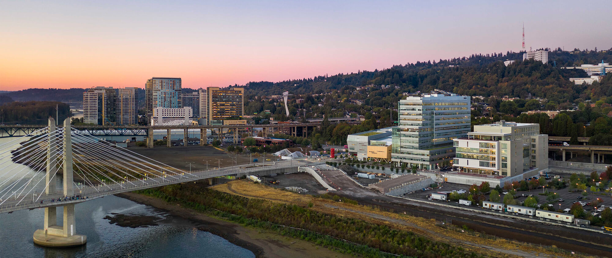
Our lab works from Portland's South Waterfront, where OHSU's Knight Cancer Institute brings together cancer biology, early detection, quantitative science, engineering, and translational medicine along the Willamette River.
Thank you to our funders and institutional partners.
This work is made possible by support for collaborative cancer research, live-cell imaging, computational modeling, early detection, and translational discovery.

Our long-term goal is to define the rules that govern tumor progression and use them to identify better points of intervention.